|
|
Carcinoma of the Gallbladder
General Considerations
- Uncommon adenocarcinoma affecting patients in their 60’s or older with long-standing cholelithiasis
- Female predominance
- Associated with
- Cholelithiasis or choledocholithiasis in 70-90% (most often cholesterol stones)
- Calcification of the wall (Porcelain gallbladder)

- Carcinoma occurs in 25% of porcelain gallbladders
- Congenital biliary cysts
- Chronic Salmonella infections
- Anomalous junction of the pancreatic and biliary ducts
- Smoking
- More common in Native Americans
Clinical Findings
- Symptoms are frequently delayed until tumor has grown and/or spread
- Pain
- Anorexia
- Nausea or vomiting
- Obstructive jaundice
- Duodenal obstruction
- Hepatomegaly
- Ascites
- Rarely, extra-abdominal metastases
Imaging Findings
- Most tumors occur in the fundus (60%), then body (30%)
- US is the study of first choice
- Necrotic extraluminal mass extending to liver is most common presentation
- Focal or diffuse thickening of the GB wall
- Intraluminal mass without shadowing
- Gallstones
- GB may be small in size, normal or distended
- Thickening of GB wall
- GB mass that may replace the entire lumen
- May enhance heterogeneously
- Gallstones
- Hepatic metastases
- Biliary dilatation
Differential Diagnosis
- Contracted gallbladder with calcified wall can be mistaken for a gallstone
- Hepatocellular carcinoma
- Cholangiocarcinoma
- Metastatic disease to gallbladder fossa
- Thickening of the wall can also occur in cholecystitis, cirrhosis, congestive heart failure, hypoalbuminemia, adenomyomatosis and hepatitis
Treatment
- Surgical resection, if possible
Complications
- Biliary obstruction
- Fistula to GI tract
Prognosis
- Lymphatic spread to retroperitoneal, right celiac, and pancreaticoduodenal nodes
- Direct invasion of the liver, extrahepatic biliary ducts, duodenum and colon
- Intraperitoneal seeding
- Can be reasonably good if resected early but lack of early symptoms frequently delays diagnosis and leads to mean survival rate of 6 months
- Found incidentally in 1-3% of cholecystectomy specimens

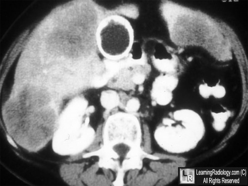
Carcinoma of the gallbladder with liver metastases. Contrast-enhanced axial CT image of the mid-abdomen demonstrates a thickened, very dense and irregular gallbladder wall (white arrow). The density of the gallbladder wall is due to calcification of the wall (porcelain gallbladder). There are low attenuation lesions in the right and left lobes of the liver (red circles) representing metastatic deposits.
For more information, click on the link if you see this icon 
For this same photo without the annotations, click here
Gallbladder Carcinoma eMedicine Szarnecki, G; Karol, G; Khalil, H
|
|
|

)